
Radiography comprises a dedicated team of radiographers & radiography assistants. We work together with radiologists and nurses to provide high-quality medical imaging services to patients in a safe and efficient manner. In addition to routine outpatient imaging services, we also provide 24-hour imaging coverage for inpatients and the Emergency department.
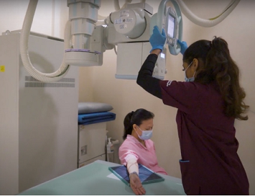
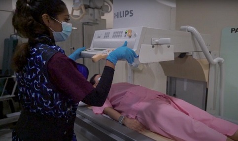
General radiography or X-rays is the most basic form of diagnostic imaging. X-rays are a form of electromagnetic radiation that can be used to create images of the human body. Our radiographers perform x-ray studies for a wide variety of conditions.
General radiography services include plain film x-ray imaging, fluoroscopy, bone mineral densitometry (BMD). We also support the needs of intraoperative imaging within the operating theaters, at the endoscopy center and during extracorporeal shock wave lithotripsy (ESWL) cases.
When doing an X-ray, there will be exposure to radiation. However, the dose of radiation is usually low and, in most cases, equivalent to a few days of radiation exposure from naturally occurring sources such as the sun or taking an airplane flight.
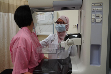
Breast imaging is the use of mammogram and ultrasound to detect for any breast abnormalities, such as breast lumps and microcalcifications, which may be cancerous. Mammograms are typically performed on women aged 40 years and above as a screening method for breast cancer, whereas breast ultrasound can be performed for women at any age. The radiographers trained in breast imaging also assist the Radiologist during breast biopsies to confirm whether a breast abnormality is cancerous.
Although x-ray (ionising radiation) is used in mammography, the dose is relatively low and safe. Nevertheless, pregnant patients and patients who are breast feeding are required to inform the radiographer before the mammogram.
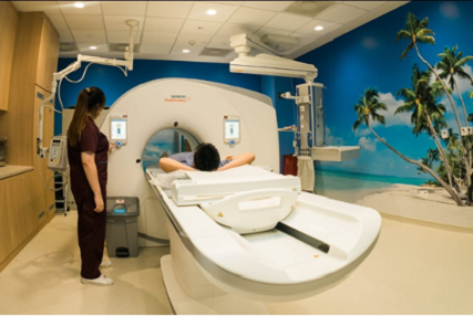
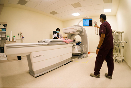
Computed Tomography (CT) scan is a diagnostic imaging procedure that uses a combination of ionising radiation and computer reconstruction technology to produce detailed images of any part of the body, including the bones, muscles, fat, organs, and blood vessels. As CT scans capture 3D information, it provides more details compared to a standard x-ray film. Some conditions require CT scans with contrast, a special dye that is injected into the veins. This is for better visualization of the organs to aid diagnosis.
With technological advancements, it is possible to keep radiation doses as low as possible. Pregnant patients are not advised to go for CT scans due to the radiation, but it may be necessary for life-threatening cases where the benefits outweigh the risks.
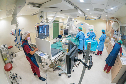
In Interventional Radiography (IR), the radiographers, nurses and interventional radiologists work seamlessly to perform minimally invasive image-guided procedures to insert vascular lines, biopsy and treat tumors and bleeding. Ultrasound and CT can be used to guide the placement of special probes which can burn tumors to kill cancer cells (i.e., tumor ablation). They can also use real time x-rays (i.e. fluoroscopy) together with contrast dye to insert wire coils into vessels to stop bleeding.
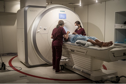
MRI is a non-invasive medical imaging test that can produce detailed 3D images of almost every internal structure in the human body, including organs, bones, muscles, and blood vessels, using a large magnet and radio waves. It offers clear imaging of soft tissue structures and is often preferred for the diagnosis of neurological conditions and spinal or central nervous system (CNS) problems. Some MRI scans may require contrast (dye) to be injected into the veins to highlight certain structures.
There is no risk of exposure to ionizing radiation as x-rays are not used during the MRI scan. However, as there is a strong magnetic field, metallic objects cannot be allowed inside the scan room. As a safety precaution, a detailed patient screening checklist will be carried out prior to the scan, to check whether they have any metallic implants, pacemaker, or other surgical clips.
Nuclear medicine imaging is a method of producing images by detecting radiation from different parts of the body after a radioactive tracer (i.e. radiopharmaceutical) is given to the patient. The images are digitally generated on a computer. Radioactive tracers used in nuclear medicine are, in most cases, injected into a vein. For some studies, they may be given orally. The amount of radiation a patient receives in a typical nuclear medicine scan tends to be very low due to short radioisotope half-life and the dose of the radioactive tracer used.
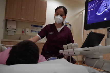
During an ultrasound examination, high frequency sound waves are transmitted into the body through a scanning probe. This allows the ultrasound machine to produce live real-time images of the internal organs. Ultrasound is commonly used to scan the organs and blood vessels in the body to detect for abnormalities. It can also assess abnormalities in the muscles, tendons, and ligaments dynamically.
As no radiation is involved, ultrasound is one of the safest advanced imaging techniques.
Mon - Thu: 8.00am - 5.30pm
Fri: 8.00am - 5.00pm
Sat, Sun & PH: Closed
(Opening Hours for Specialist Outpatient Clinic Level 5: Mon-Fri 8.30am - 6pm)
Ng Teng Fong General Hospital
Tower A - Specialist Outpatient Clinics
Level 1
Mon - Thu: 8.00am -5.30pm
Fri: 8.00am - 5.00pm
Sat: 8.00am - 12.00pm
Sun & PH: Closed
(24/7 service for X-ray at Radiology Level 1 & ED, OT, CVL and CT ED)
Ng Teng Fong General Hospital
Tower B - Ward
Level 1